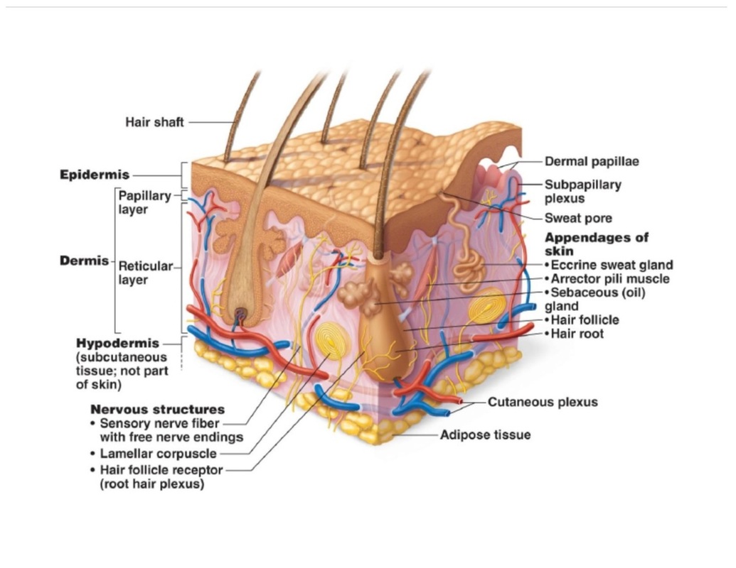
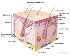
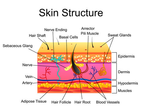
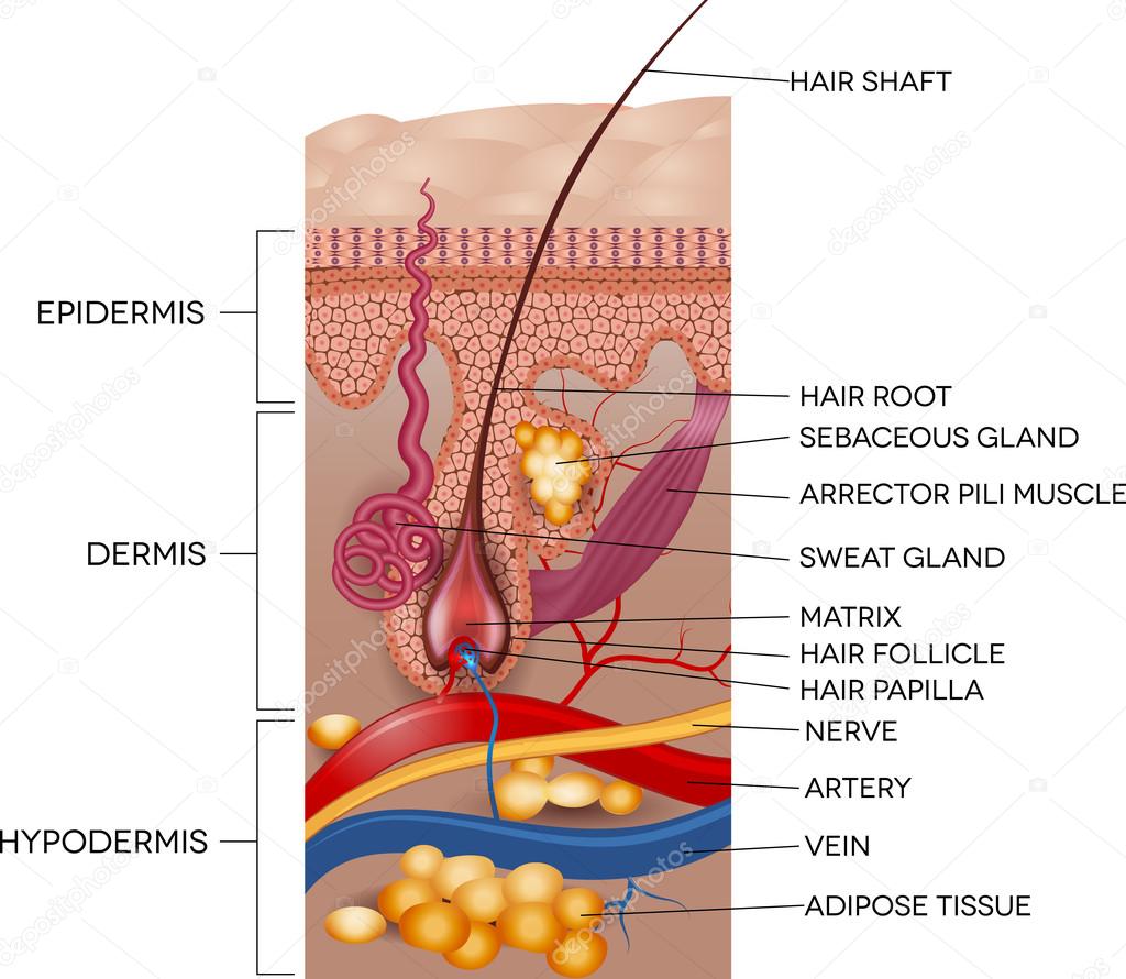
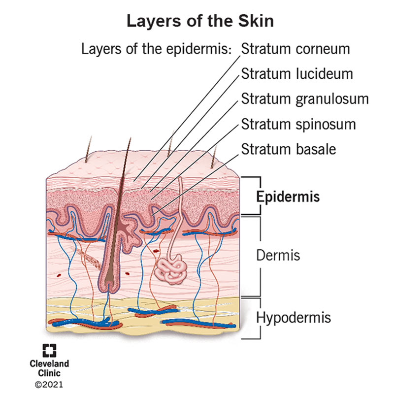
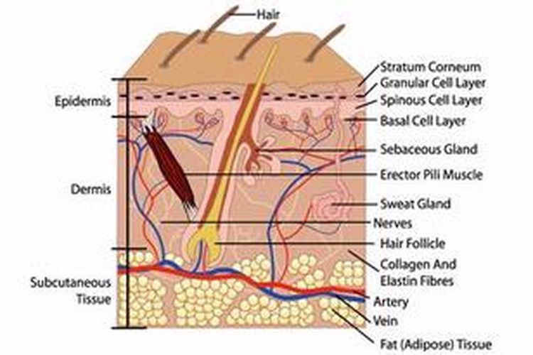
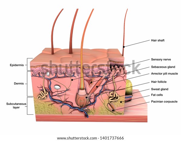
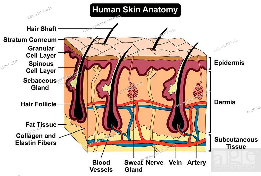
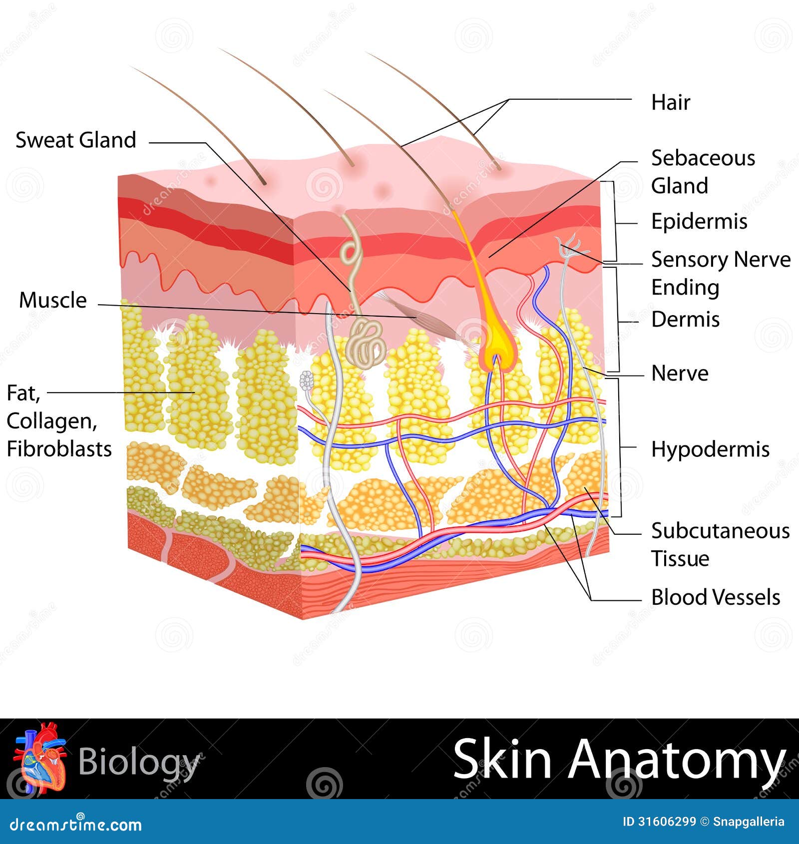
38 skin diagram with labels
Box on an inclined plane free body diagram Il y a 2 jours · The inclined plane makes an angle of 220 with the horizontal plane.When the box is moving on the inclined Plane, there is a friction force Ff of 5 N opposing the motion.The box was Originally at a height h from the ground (h=4 m). Draw the free body diagram for the box.Determine the components x, y for all the forces acting on the box.A sphere or cylinder on … Label Skin Diagram Printout - EnchantedLearning.com Read the definitions, then label the skin anatomy diagram below. blood vessels - Tubes that carry blood as it circulates. Arteries bring oxygenated blood from the heart and lungs; veins return oxygen-depleted blood back to the heart and lungs. dermis - (also called the cutis) the layer of the skin just beneath the epidermis.
How to draw skin LS - Pinterest Apr 13, 2016 - Step by step tutorials on drawing biology diagrams. Easy ways of drawing biology figures.

Skin diagram with labels
Science A-Z Science Diagrams - Visual Teaching Tools Science Diagrams from Science A-Z provide colorful, full-page models of important, sometimes complex science concepts. Science Diagrams, available in both printable and projectable formats, serve as instructional tools that help students read and interpret visual devices, an important skill in STEM fields. Skin Diagram To Label Illustrations, Royalty-Free Vector ... - iStock Browse 91 skin diagram to label stock illustrations and vector graphics available royalty-free, or start a new search to explore more great stock images and vector art. Newest results tissues of the body affected by autoimmune attack tissues of the body affected by autoimmune attack. Vector illustration for medical use Leg vector icon. Simple Skin Diagram Simple Skin Diagram. Tornado draw step drawing storm simple realistic. Cell membrane. Cell labeled onion epidermal elodea leaf cells labels lab function diagram microscope label salt every manual following slide visible living Simple Skin Diagram. Free Skull Tattoo Stencils, Download Free Skull Tattoo Stencils png we have 8 Images about Free ...
Skin diagram with labels. Skin diagram labeled - Healthiack Skin diagram labeled This brief article displays Skin diagram labeled … Please click on the diagram (s) to view larger version. You are welcome to browse healthiack.com for more details on this very topic. Best viewed on 1280 x 768 px resolution in any modern browser. Skin diagram labeled 1075 Skin diagram labeled 1077 Skin diagram labeled 1080 Peanut and Nut Allergies: Common Foods, Items to ... - WebMD Skin rash; Runny nose; Tingling in your mouth or throat, ... Nix them when you cook, and look for them on food labels: Nut butters: Almond, cashew, peanut, and others; Nut pastes. These include ... Layers of Skin: How Many, Diagram, Model, Anatomy, In Order - Healthline The epidermis is the top layer of your skin. It's the only layer that is visible to the eyes. The epidermis is thicker than you might expect and has five sublayers. Your epidermis is constantly... Anatomy of the Skin - Stanford Children's Health The skin is made up of 3 layers. Each layer has certain functions: Epidermis. Dermis. Subcutaneous fat layer (hypodermis) ...
Skin 1: the structure and functions of the skin - Nursing Times by S Lawton · 2019 · Cited by 45 — Stratum basale (germinative layer). Fig-2-Layers-of-the-skin-1024x758.jpg. The epidermis also contains other cell structures. Keratinocytes make ... 654 Skin Layers Diagram Premium High Res Photos - Getty Images 654 Skin Layers Diagram Premium High Res Photos Browse 654 skin layers diagram stock photos and images available, or search for skin cells to find more great stock photos and pictures. of 11 NEXT Human Heart (Anatomy): Diagram, Function, Chambers, Location in ... - WebMD WebMD's Heart Anatomy Page provides a detailed image of the heart and provides information on heart conditions, tests, and treatments. Skin diagram - Teaching resources - Wordwall layers of the skin Labelled diagram by Emmawebster Structure of the skin Labelled diagram by Lfoster1 Reflection diagram Labelled diagram by Elizabeth312 KS3 Physics Skin A&P Labelled diagram by Laurenbutler Adult Education Structures of the skin Labelled diagram by Lindabiles Hair and Skin Labelled diagram by Michaela131
label the skin diagram - Printable - PurposeGames.com This is a free printable worksheet in PDF format and holds a printable version of the quiz label the skin diagram. By printing out this quiz and taking it with pen and paper creates for a good variation to only playing it online. Simple diagram of the skin - Healthiack Best viewed on 1280 x 768 px resolution in any modern browser. Simple diagram of the skin 1285. Simple diagram of the skin 1286. Simple diagram of the skin 1296. Simple diagram of the skin 1300. Simple diagram of the skin 1302. Simple diagram of the skin 1304. Simple diagram of the skin 1308. Simple diagram of the skin 1311. Labeled Skin Structure Diagram | Quizlet skin structure. ... Hair Shaft. Nonliving, extracellular matrix produced and secreted by hair follicle cells. Involved in protection, sensation, and temperature regulation. Epidermis. Outermost layer of skin, provides a strong, waterproof, protective barrier for the body. Dermis. Fibrous and elastic tissue, provides strength and elasticity to ... Skin Diagram || How to draw and label the parts of skin It contains cutaneous receptors for touch. The skin consists of two main layers called epidermis and dermis. Epidermis is the layer of protection. It has sweat pores and small hairs. Dermis lies...
Skin Labeling Quiz - PurposeGames.com This is an online quiz called Skin Labeling. There is a printable worksheet available for download here so you can take the quiz with pen and paper. From the quiz author. Epidermis, Dermis, Hypodermis Your Skills & Rank. Total Points. 0. Get started! Today's Rank--0. Today 's Points. One of us!
skin labeling Diagram | Quizlet Epidermis. outermost layer of the skin;composed of squamous epithelium; contains keratin. subcutaneous layer. Innermost layer of skin, contains fat and is the location of main blood vessels. basal layer of epidermis. The deepest layer of the Epidermis (outermost layer of the skin). The cells in the basal layer are alive, multiplying and growing.
Skin Histology Slide Identification - AnatomyLearner Let's identify the thick and thin skin histology slides under a light microscope. First, talk about the thin skin microscope slide identification. #1. The provided tissue section shows two distinct layers - the epidermis and dermis. #2. Presence of thin epidermis that lines with keratinized stratified squamous epithelium. #3.
A physical wiring diagram for the human immune system | Hacker … 08/08/2022 · A physical wiring diagram for the human immune system (nature.com) 169 points by bookofjoe 9 hours ago | hide | past | favorite | 23 comments: photochemsyn 6 hours ago | next. This is kind of stunning. They appear to have discovered something like 30 novel (previously unknown) immune cell receptor interactions (Fig 1). Then they built a quantitive model, looked …
Skin diagram to label - Labelled diagram - Wordwall Skin diagram to label - Labelled diagram Epidermis, Dermis, Hypodermis, Blood and lymph, Sensory nerve ending, Sweat gland, Arrector pili muscle, Sebaceous gland, Hair shaft, Dermal papilla, Hair follicle. Skin diagram to label Share by Kmiller14 Like Edit Content More Leaderboard Theme Options Switch template Interactives
Basic Skin Diagram Basic Skin Diagram. Basic principles in plastic surgery pptx. Image result for hair follicle model labeled. Liver cell drawing diagram draw label ultrastructure paintingvalley animal example labelling diagrams ze kig source. Basic Skin Diagram. Basic principles in plastic surgery pptx - د. ياسر نايف - Muhadharaty we have 9 Pictures ...
skin anatomy diagram labeled - Microsoft Skeleton human system skeletal body labels bones labeled anatomy 206 digestive born many enchantedlearning humans parts label bone structure function. Sectioned sebaceous. Skin structure game quiz purposegames statistics games skin anatomy diagram labeled
Skin Diagram with Detailed Illustrations and Clear Labels - BYJUS Skin Diagram. The largest organ in the human body is the skin, covering a total area of about 1.8 square meters. The skin is tasked with protecting our body from external elements as well as microbes. Interesting Note: The skin is also responsible for maintaining our body temperature – this was apparent in victims who were subjected to the medieval torture of being skinned alive. …
Consumer Updates | FDA - U.S. Food and Drug Administration Jul 28, 2022 · The .gov means it’s official. Federal government websites often end in .gov or .mil. Before sharing sensitive information, make sure you're on a federal government site.
Skin Diagram | Worksheet | Education.com Skin Diagram. Though skin may seem like nothing more than the source of things like zits and oil to your pre-teen, skin actually has many important jobs to do. Learn more about the skin (and the science behind pimples -- ew!) in this printable life science diagram.
Diagram Of Body Organs Female Pics Stock Photos, Pictures ... Human skin changes or ageing skin. A diagram of younger and older skin showing the decrease in collagen fibers, atrophy and broken elastin, formed wrinkles, hair becomes gray in the elderly. Multiple sclerosis anatomical vector illustration diagram, medical scheme. Multiple sclerosis anatomical vector illustration diagram, medical scheme with affected area icons and damaged …
Labeled Skin Diagram Pictures, Images and Stock Photos Labeled medical diagram, a 3D cross section of human skin layers and parts such as a hair follicle and sweat glands on a white background. Sensory receptors in the human skin Cutaneous Receptors. Sensory receptors in the human skin. labeled. Human anatomy Exfoliation. cosmetology Exfoliation. mechanical means. cosmetology.
Skin Anatomy: The Layers of Skin and Their Functions - Verywell Health The epidermis is the outermost layer of the skin. Its thickness depends on where it is located on the body. It is thinnest on the eyelids (roughly half a millimeter) and thickest on the palms and soles (1.5 millimeters). The epidermis is made up of five individual layers: 2
Object Diagram syntax and features - PlantUML.com Object08 : some labels @enduml Associations objects. Edit in Dokuwiki Edit in Asciidoc Edit in Markdown. 🎉 Copied! @startuml object o1 object o2 diamond dia object o3 o1 --> dia o2 --> dia dia --> o3 @enduml Adding fields. Edit in Dokuwiki Edit in Asciidoc Edit in Markdown. To declare fields, you can use the symbol : followed by the field's name. 🎉 Copied! @startuml object user user ...
Pin by Benjamin Brown on Pathogenicity | Skin anatomy, Human anatomy ... This article will look at the components and the accessory structures of the integumentary system, skin healing, skin integrity, and the staging of pressures ulcers. This article contains 7 Facts about the Integumentary System Every Nursing Student Should Know. #nursecepts #iintegumentarysystem #nursingstudent #nursingschool. N.
Skin Labeling | Biology Game | Turtle Diary Skin Labeling - Biology Game Identify and label figures in Turtle Diary's interactive online game, Skin Labeling! Drag the given words to the correct blanks to complete the labeling!
Diagram of human skin structure — Science Learning Hub Diagram of human skin structure. Image. Add to collection. Tweet. Rights: University of Waikato Published 1 February 2011 Size: 100 KB Referencing Hub media. The epidermis is a tough coating formed from overlapping layers of dead skin cells.
Label the skin - Teaching resources - Wordwall Label the skin - Label the skin - Label the diagram of the skin - Skin diagram to label - The Skin - Skin diagram to label - The Skin - The skin.
Picture of the Skin - WebMD The skin is the largest organ of the body, with a total area of about 20 square feet. The skin protects us from microbes and the elements, helps regulate body temperature, and permits the...
Male Human Anatomy Diagram Pictures, Images and Stock Photos Male and female body chart Male and female body chart, front and back view. Blank human body template for medical infographic. Isolated vector clip art illustration. male human anatomy diagram stock illustrations
Skin Diagram Labeling Skin Diagram Labeling. 1. Label the diagram with the letters below according to the structure/area they describe. You may label with a line or put the label ...
skin diagram to label | Anatomy coloring book, Body systems worksheets ... skin diagram to label. Find this Pin and more on Homeschool by Stephanie Parker. Science Topics. Science Biology. Anatomy Coloring Book. Coloring Books. Human Body Activities. Skin Facts. Integumentary System.
Skin Diagram Teaching Resources | Teachers Pay Teachers Integumentary System: Skin Diagram to Label by Lori Maldonado 4.9 (30) $2.00 PDF Students will read the definitions and label the skin anatomy diagram. Answer key included. This diagram had been modified from Enchanted Learning. I have used this worksheet as an in-class assignment as well as a homework assignment.
PDF Skin Diagram Labeling - New Providence School District Skin Diagram Labeling . 1. Label the diagram with the . letters. below according to the structure/area they describe. You may label with a line or put the label directly onto the area described. Be as precise as possible. If you are worried about the precision of your label add a word after to explain exactly where your label should be.
Anatomy, Diagram and Function of Skin - VEDANTU The skin has a surface area of between 16.1-21.5 sq ft. for an adult human. The thickness of the skin differs over all parts of the body, and between men and women and the young and the old. For example, the skin on the forearm which is on average 1.3 mm in the human male and 1.26 mm in the human female.
Simple Skin Diagram Simple Skin Diagram. Tornado draw step drawing storm simple realistic. Cell membrane. Cell labeled onion epidermal elodea leaf cells labels lab function diagram microscope label salt every manual following slide visible living Simple Skin Diagram. Free Skull Tattoo Stencils, Download Free Skull Tattoo Stencils png we have 8 Images about Free ...
Skin Diagram To Label Illustrations, Royalty-Free Vector ... - iStock Browse 91 skin diagram to label stock illustrations and vector graphics available royalty-free, or start a new search to explore more great stock images and vector art. Newest results tissues of the body affected by autoimmune attack tissues of the body affected by autoimmune attack. Vector illustration for medical use Leg vector icon.
Science A-Z Science Diagrams - Visual Teaching Tools Science Diagrams from Science A-Z provide colorful, full-page models of important, sometimes complex science concepts. Science Diagrams, available in both printable and projectable formats, serve as instructional tools that help students read and interpret visual devices, an important skill in STEM fields.
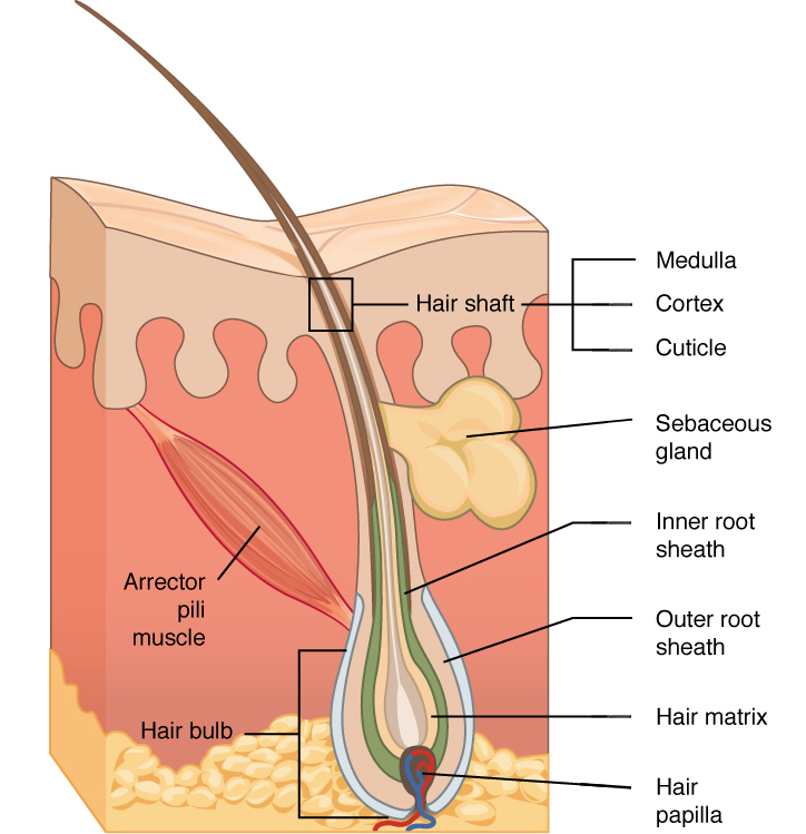

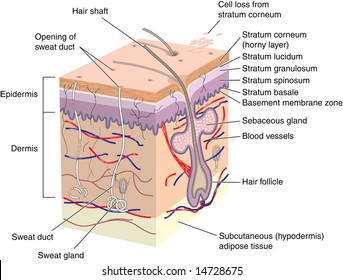
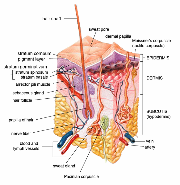


:background_color(FFFFFF):format(jpeg)/images/library/11027/labeled_diagram_anatomy_of_integumentary_system.jpg)


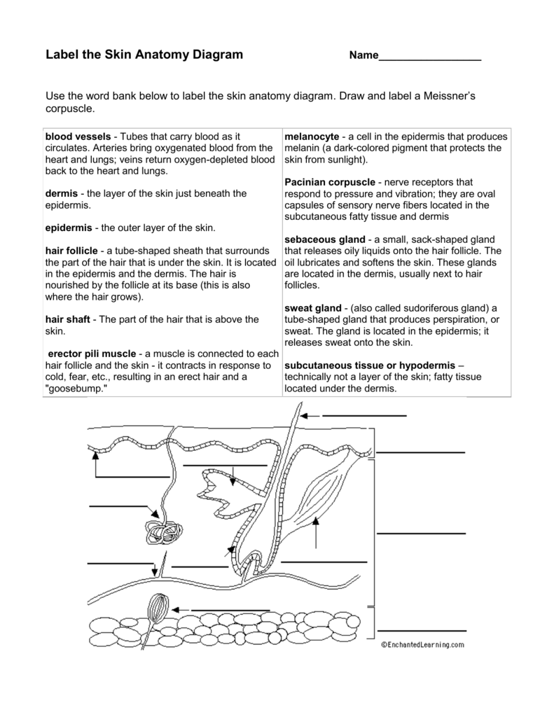








Post a Comment for "38 skin diagram with labels"