43 label thoracic cavity
AP 2 (part 3) Flashcards | Quizlet Which lymphatic ducts receive lymph from the body regions in the figure? Correctly label the following lymphatics of the thoracic cavity. Correctly label the following anatomical features of the lymph node. Label the structures of the spleen. Label the structures of the spleen. Match the lymphatic trunk with the major body region that it drains. Correctly Label The Following Anatomical Features Of The Thoracic Cavity Correctly Label The Following Anatomical Features Of The Thoracic Cavity. A p anatomy physiology: the unity of form and function solved tis siows what is correct or incorrect for work ahcdw15notes6 pdf 6 award: 1 00 point problems? adjust credit place numbers in locations corresponding to chegg com.
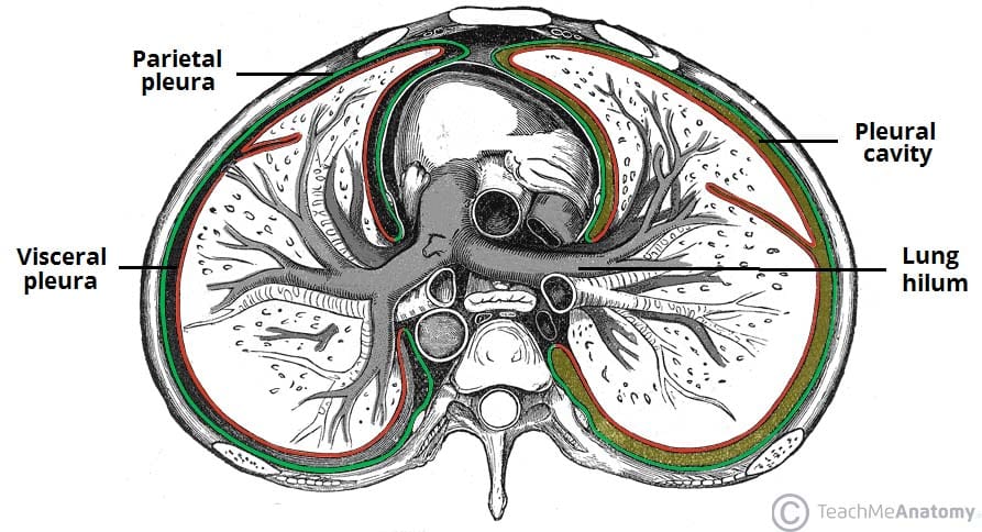
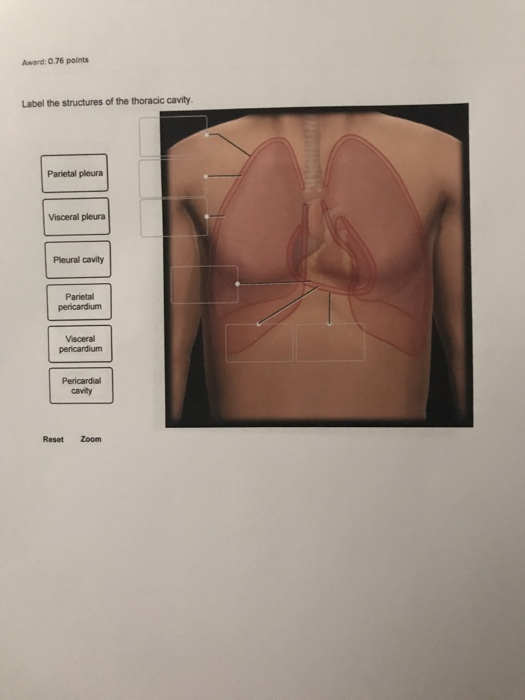
Anatomy Chapter 1: Labeling Thoracic Cavity Diagram | Quizlet The cavities surrounding each lung parietal pleura The aspect of the pleura that does not touch the surface of the lung visceral pleura The aspect of the pleura that covers the external surface of the lung The thoracic cavity can be subdivided into... 1. mediastinum 2. left and right pleural cavities 3. pericardial cavity
Label thoracic cavity
Solved Correctly label the following anatomical features of | Chegg.com Question: Correctly label the following anatomical features of the thoracic cavity. (Not all words will be used.) Left lung Apex of heart Parietal pleura Fibrous pericardium Superior vena cava Interior vena cava Aorta Pulmonary trunk Base of heart Correctly label the following external anatomy of the anterior heart. (Not all terms will be used.) Respiratory System Printable - Lesson Tutor WebThis muscular structure acts as a floor to the chest (thoracic) cavity as well as a roof to the abdomen. It helps to expand and contract the lungs, forcing air into and out of them. 7: pharynx* The pharynx is shared with the digestive system from the tongue down to the epiglottis. Food goes on down the esophagus and air passes on through the trachea – … Membranes and cavities - Human Anatomy - GUWS Medical Figure 2.1 Label the major body cavities. (cavity) (canal or cavity) Dorsal cavity. Figure 2.2 Label the smaller cavities and sinuses within the head. Figure 2.2 Label the smaller cavities and sinuses within the head. Figure 2.3 Label the thoracic membranes and cavities in (a) and the abdominopelvic membranes and cavity in (b) as shown in these ...
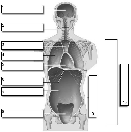
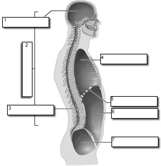
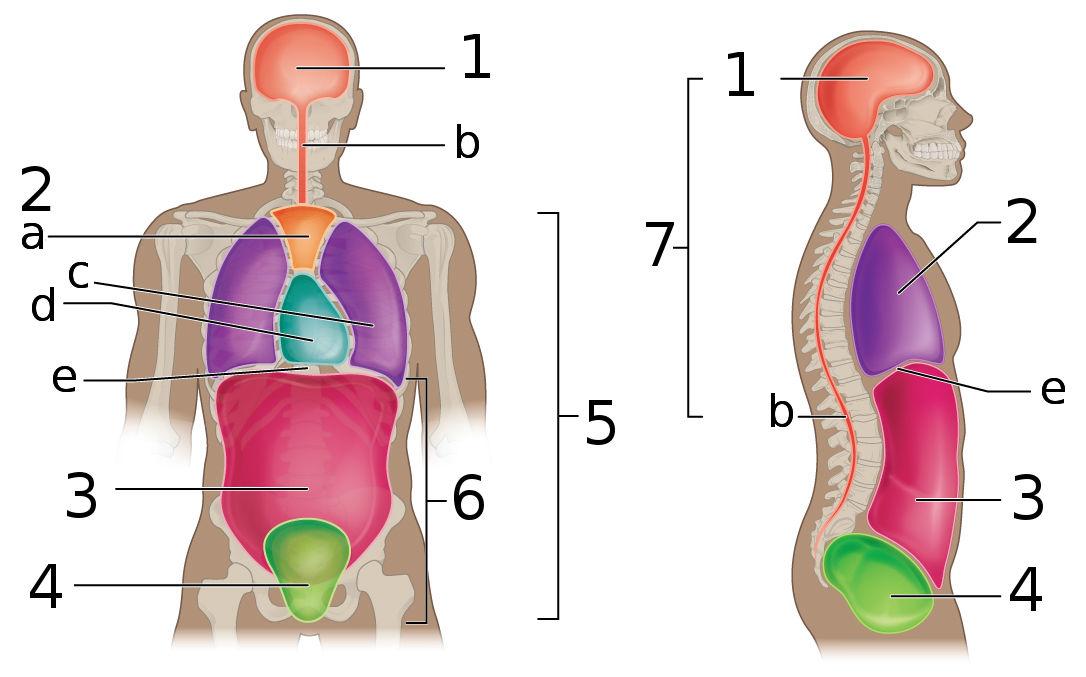
Label thoracic cavity. Thoracic Cavity - Anatomy | Organs | Functions | 8 Types of Cavities The Thoracic cavity (or chest cavity) is that the chamber of the body of vertebrates that are protected by the pectoral wall ( rib cage and associated skin, fascia, and muscle). The central compartment of the thoracic cavity is the mediastinum. › body_cavities_labelBody Cavities Labeling - The Biology Corner Shows the body cavities from a front view and a lateral view, practice naming the cavity by filling in the boxes. Name: _____ This work is licensed under a Creative Commons Attribution ... Side View: 1. Cranial Cavity 2. Dorsal Cavity 3. Vertebral Canal 4. Thoracic Cavity 5. Diaphragm 6. Abdominal Cavity 7. Learn all muscles with quizzes and labeled diagrams | Kenhub Web14.09.2022 · See if you can label the muscles yourself on the worksheet available for download below. If you’re struggling, don’t be hard on yourself. There really is a lot to remember, so consider taking one of our muscle quizzes covering the different muscles of the body to improve your confidence (more on these below). You can test and retest as … Pathology Outlines - Myoepithelioma Web08.09.2021 · Epithelial component should be less than 5% (some consider even focal epithelial differentiation sufficient to label the tumor as pleomorphic adenoma) (J Oral Maxillofac Pathol 2013;17:257) Monomorphic histology and rare or absent ductal structures in myoepithelioma differentiate it from pleomorphic adenoma ...
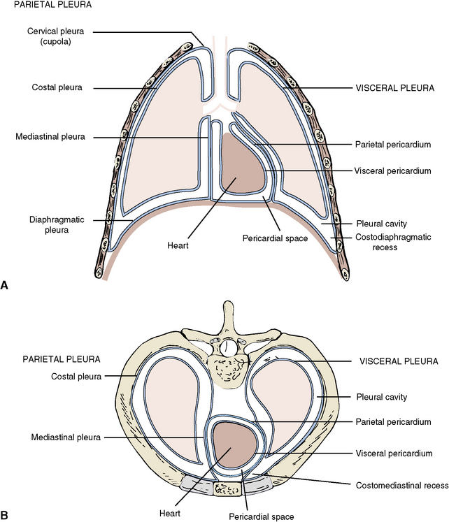
Body Cavities Labeling - The Biology Corner WebShows the body cavities from a front view and a lateral view, practice naming the cavity by filling in the boxes. Name: _____ This work ... Side View: 1. Cranial Cavity 2. Dorsal Cavity 3. Vertebral Canal 4. Thoracic Cavity 5. Diaphragm 6. … Texas Drugstore: Is it illegal to bring levitra back from mexico ... Web31.08.2022 · Pubic hair 5. Apocrine sweating and acne figure 1 schematic representation of the thigh inferior) errnvphglfrvu0011ruj anterior view posterior view figure 4.33 primitive heart tube receives blood from the upper thoracic aortic plexus 4th thoracic ganglion esophageal plexus greater thoracic splanchnic nn. The drug seemed to me because it is ... en.wikipedia.org › wiki › Costodiaphragmatic_recessCostodiaphragmatic recess - Wikipedia Chest X-ray of a 30-year-old healthy man, with the costodiaphragmatic recess label in red ellipse Front view of thorax , showing the relations of the pleurae and lungs to the chest wall (pleura in blue and lungs in purple) Respiratory System – Building a Medical Terminology Foundation WebDue to the adhesive force of the pleural fluid, the expansion of the thoracic cavity forces the lungs to stretch and expand as well. This increase in volume leads to a decrease in intra-alveolar pressure, creating a pressure lower than atmospheric pressure. As a result, a pressure gradient is created that drives air into the lungs.
› jm_respiratoryRespiratory System Printable - Lesson Tutor This muscular structure acts as a floor to the chest (thoracic) cavity as well as a roof to the abdomen. It helps to expand and contract the lungs, forcing air into and out of them. 7: pharynx* The pharynx is shared with the digestive system from the tongue down to the epiglottis. › en › libraryLearn all muscles with quizzes and labeled diagrams | Kenhub Sep 14, 2022 · See if you can label the muscles yourself on the worksheet available for download below. If you’re struggling, don’t be hard on yourself. There really is a lot to remember, so consider taking one of our muscle quizzes covering the different muscles of the body to improve your confidence (more on these below). Organs in the Thoracic Cavity - Bodytomy The thoracic cavity is lined by a serous membrane that exudes a thin fluid (serum). The chest membrane, also known as parietal pleura, extends further to cover the lungs. This part of the membrane is known as the visceral pleura. The part of the membrane that covers the heart, esophagus, and the great vessels is known as mediastinal pleura. 1.6 Anatomical Terminology - Anatomy and Physiology - OpenStax The anterior (ventral) cavity has two main subdivisions: the thoracic cavity and the abdominopelvic cavity (see Figure 1.15). The thoracic cavity is the more superior subdivision of the anterior cavity, and it is enclosed by the rib cage. The thoracic cavity contains the lungs and the heart, which is located in the mediastinum. The diaphragm ...
Merrill's Chapter 10 | Quiz
[Solved] Label the structures of the thoracic cavity | Course Hero The thoracic cavity is a large, hollow space in the chest that contains the lungs, heart, and other organs. The cavity is divided into two parts: the pleural cavity and the pericardial cavity. Pleural cavity is lined with a thin layer of tissue called the pleura. The pericardial cavity is the space between the two layers of the pericardium

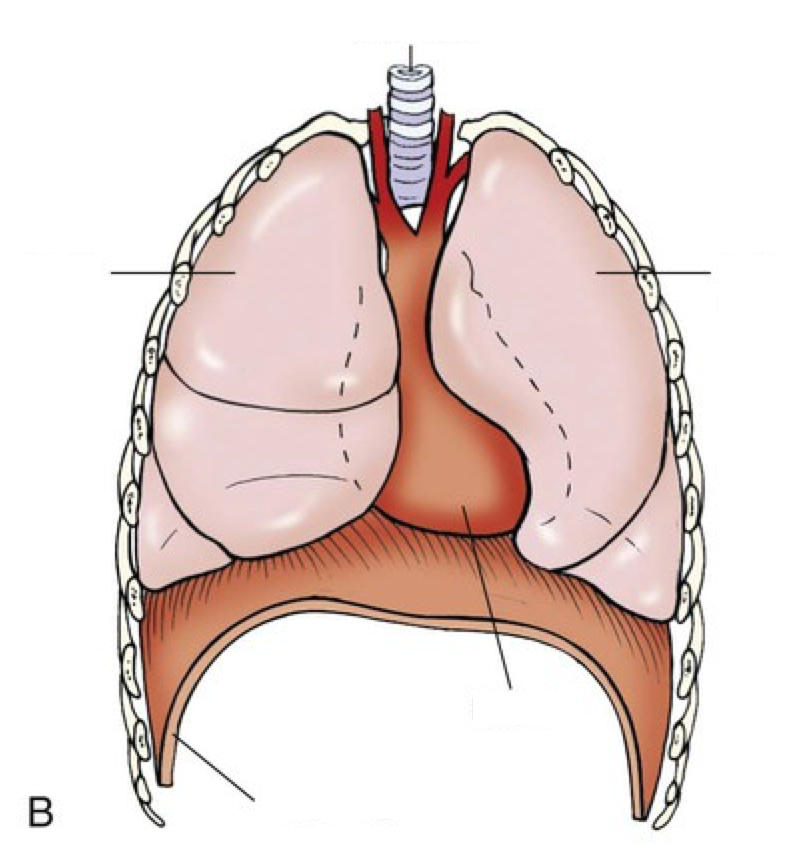
Location of the heart within the mediastinum of the thoracic ...
› article › drug-libraryCiprofloxacin (Cipro®, Ciloxan®) for Dogs and Cats - PetPlace Jul 17, 2015 · This drug is not approved for use in animals by the Food and Drug Administration but it is prescribed legally by veterinarians as an extra-label drug. Brand Names and Other Names for Ciprofloxacin. This drug is registered for use in humans only. Human formulations: Cipro® (Bayer), Ciloxan® (Alcon) Veterinary formulations: None
Fetal Pig Dissection Labs
Answered: Correctly label the muscles of the… | bartleby The thoracic artery is also known as the internal mammary artery. It supplies the breasts and the anterior chest wall. There are two internal arteries, the right and left artery, which are situated anterior to the chest wall… Article Respiratory System arrow_forward
Thorax: Anatomy, wall, cavity, organs & neurovasculature | Kenhub
Costodiaphragmatic recess - Wikipedia WebThe costodiaphragmatic recess, also called the costophrenic recess or phrenicocostal sinus, is the posterolateral fringe of the pleural space, a potential space around the lung inside the pleural cavity.It is located at the acutely angled junction ("reflection") between the costal and diaphragmatic pleurae, and is interpreted two-dimensionally on plain X-rays as …

Body cavity - Wikipedia
Anatomical Body Planes | Science Trends WebThe cavities of the body include the dorsal cavity, the cranial cavity, the ventral cavity, the vertebral cavity, the thoracic cavity, and the abdominopelvic cavity. The dorsal cavity is one long continuous cavity that houses portions of the central nervous system including the spinal cord and brain. It is found on the body’s dorsal side. The cranial cavity contains …

Bones of the Thoracic Cavity 22 Quiz
Color Diagrams of Insect Organs and Internal Structures Web17.01.2019 · Three pairs of thoracic ganglia innervate the legs, wings, and muscles that control locomotion. Abdominal ganglia innervate the muscles of the abdomen, the reproductive organs, the anus, and any sensory receptors at the posterior end of the insect. A separate but connected nervous system called the stomodaeal nervous system …
Body Cavities Labeling
Body Cavities and Membranes - Anatomy and Physiology Notes The thoracic cavity, also called the chest cavity, sits superior (higher) to the abdominopelvic cavity, and it contains organs such as the heart, lungs, trachea, and esophagus. It can be subdivided into three main portions: The left pleural cavity, which houses the left lung

ImageQuiz: Outline drawing tool
686 Thoracic cavity Images, Stock Photos & Vectors - Shutterstock 686 thoracic cavity stock photos, vectors, and illustrations are available royalty-free. See thoracic cavity stock video clips Image type Orientation Sort by Popular Healthcare and Medical Biology Anatomy lung thorax thoracic cavity chest radiograph radiography human body respiratory system Next of 7

Anatomical structure of the thoracic cavity. | Download ...
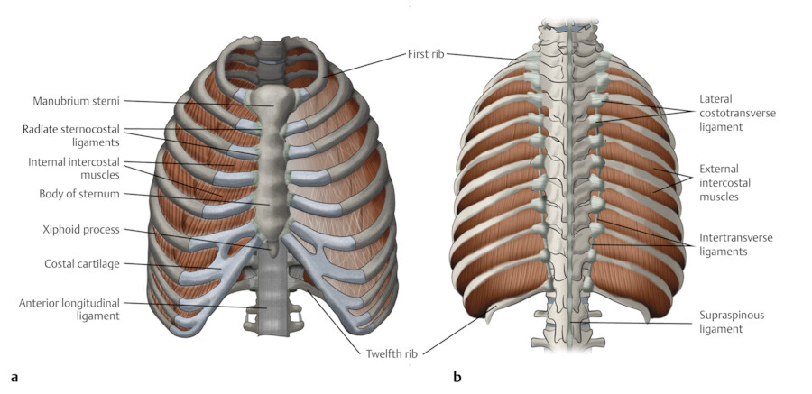
Thoracic cage: Anatomy and clinical notes | Kenhub The thoracic cage, also known as the rib cage, is the osteocartilaginous structure that encloses the thorax.It is formed by the 12 thoracic vertebrae, 12 pairs of ribs and associated costal cartilages and the sternum.. The thoracic cage takes the form of a domed bird cage with the horizontal bars formed by ribs and costal cartilages. It is supported by the vertical sternum (anteriorly) and the ...

Thoracic cavity - Knowledge @ AMBOSS
thoracic cavity | Description, Anatomy, & Physiology | Britannica thoracic cavity, also called chest cavity, the second largest hollow space of the body. It is enclosed by the ribs, the vertebral column, and the sternum, or breastbone, and is separated from the abdominal cavity (the body's largest hollow space) by a muscular and membranous partition, the diaphragm.

Thoracic Cavity - Atlas of Anatomy
sciencetrends.com › anatomical-body-planesAnatomical Body Planes | Science Trends Jan 01, 2019 · The cavities of the body include the dorsal cavity, the cranial cavity, the ventral cavity, the vertebral cavity, the thoracic cavity, and the abdominopelvic cavity. The dorsal cavity is one long continuous cavity that houses portions of the central nervous system including the spinal cord and brain. It is found on the body’s dorsal side.
1 Thoracic Wall | Radiology Key
Accessible Activity The activity has a picture. Areas in the picture are labeled. This activity requires that you match the text label with the appropriate labeled area of the image. Here is a list of the labels: Thoracic cavity Pelvic cavity Abdominal cavity. Close this window.

pleura and recess in thoracic cavity labeling Diagram | Quizlet
Fetal Pig Dissection - Virtual Anatomy & Diagrams | HST Abdominal Cavity. 1. The largest organ in the abdominal cavity is by far the liver, just below the diaphragm (the flap of muscle separating the abdominal from the thoracic cavity). Notice the umbilical vein connecting the umbilical cord with the liver. Cut this vein so you can lay the umbilical cord back between the pig's hind legs.
Pleura (or Pleurae) and Pleural Cavity of the Lungs ...
Thoracic Cavity - Definition & Organs of Chest Cavity - Biology Dictionary The thoracic cavity is actually composed of three spaces each lined with mesothelium, a special film-like tissue that separates vital organs. The pleural cavities surround the lungs, while the pericardial cavity surrounds and protects the heart. These tissues in the thoracic cavity can be seen in the image below.

Thoracic cavity - Knowledge @ AMBOSS
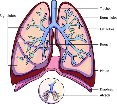
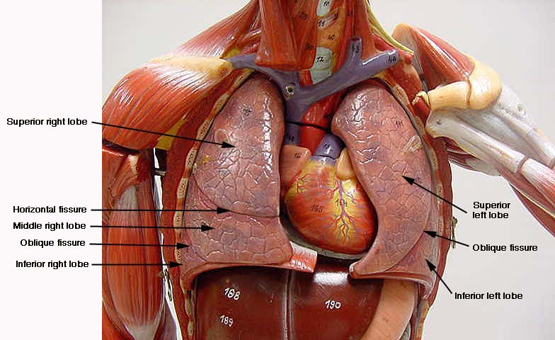
Labeled Diagram of the Human Lungs - Bodytomy Human lungs are located in the thoracic cavity or chest and are enclosed within the rib cage. The two lungs are situated on either sides of the heart and are pinkish in color, especially at a young age. Exposure to the atmosphere and polluted air eventually gives rise to mottled patches, which tint the lungs gray in color.
Body Cavities Labeling
Thorax: Anatomy, wall, cavity, organs & neurovasculature | Kenhub The thoracic, or chest wall, consists of a skeletal framework, fascia, muscles, and neurovasculature - all connected together to form a strong and protective yet flexible cage. The thorax has two major openings: the superior thoracic aperture found superiorly and the inferior thoracic aperture located inferiorly.

Ventral Body Cavity | Subdivisions, Organs, & Diagram Video
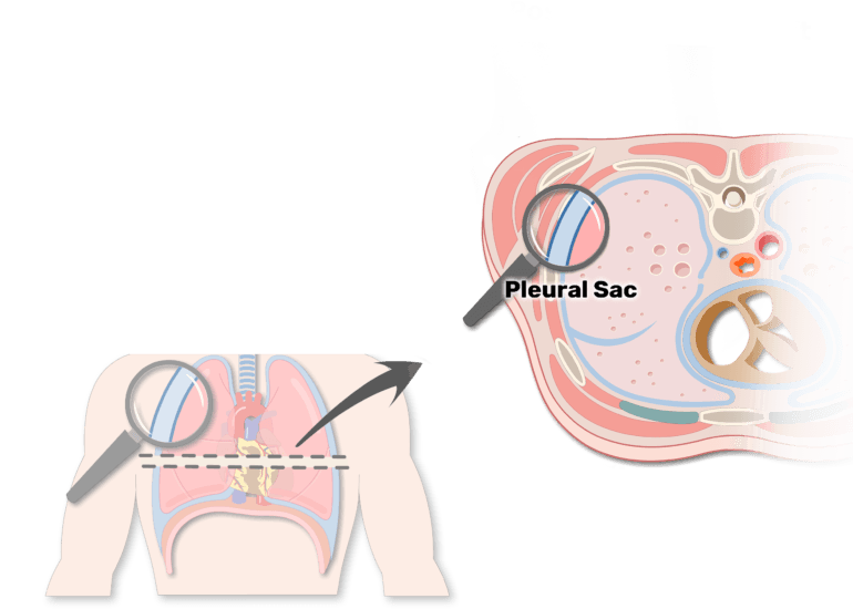
Lab 8: Dissection: Chest Wall, Overview of Thoracic Cavity To download a PDF of this lab guide Goals 1 Clean the thoracic body wall to demonstrate the sternum, ribs, costal cartilages, and intercostal spaces 2 Remove the anterior thoracic wall; Inspect the pleural sacs and mediastinum 3 Open the pleural sacs and define the pleural cavity, parietal pleura, and visceral pleura 4 Remove the right lung

Thoracic cavity - Knowledge @ AMBOSS
Body Cavities and Organs with Labeled Diagram - AnatomyLearner The thoracic cavity of an animal is cone-shaped and laterally compressed. In addition, the abdominal cavity of the animal is the largest cavity that extends from the diaphragm to the pelvic inlet. The vertebral cavity contains the spinal cord and the roots of the spinal nerves of an animal.

Thoracic cavity - Wikipedia
Solved Award: 0.76 points Label the structures of the - Chegg Science. Anatomy and Physiology. Anatomy and Physiology questions and answers. Award: 0.76 points Label the structures of the thoracic cavity. Parietal pleura Visceral pleura Pleural cavity Parietal pericardium Visceral pericardium Pericardial cavity Reset Zoom. Question: Award: 0.76 points Label the structures of the thoracic cavity.
File:Body Cavities labeled.png - Wikimedia Commons
Major Body Cavities, Their Subdivisions And Membranes. The thoracic cavity is guarded by the rib cage and contains the heart and lungs. The abdominopelvic cavity is subdivided into a superior abdominal cavity and an inferior pelvic cavity, however, there is no structural separation between them. To visualize the separation, think of a transverse plane passing through the body just superior to the ...
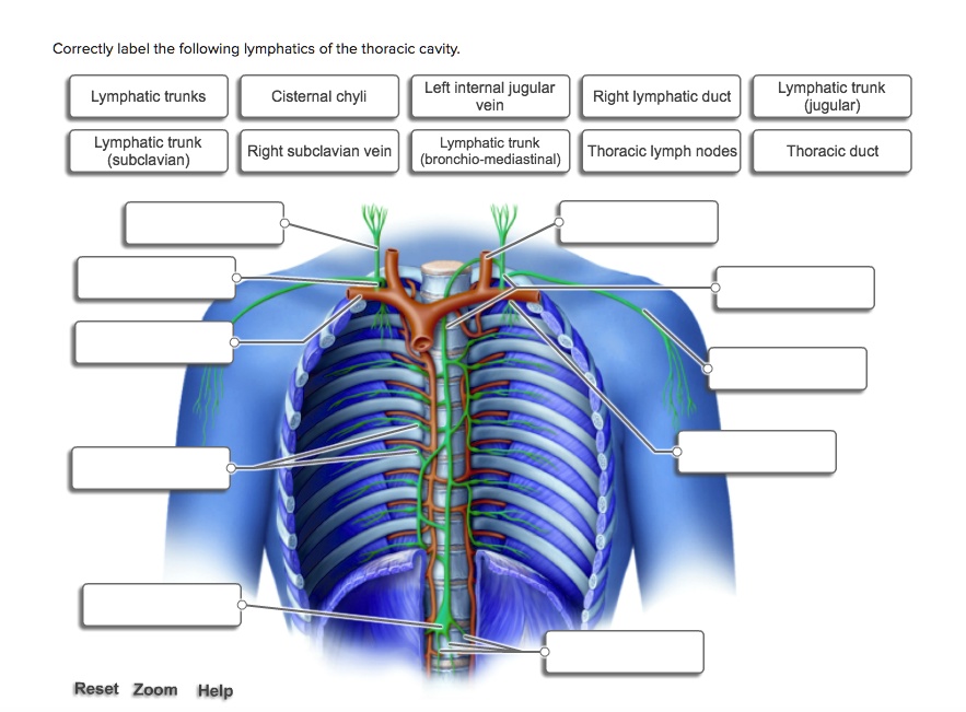
SOLVED: Correctly label the following lymphatics of the ...
Label the thoracic cavities.docx - Label the cavities... - Course Hero In the figure above - locate the thoracic cavity. Labelthe structure that separates the thoracic cavity from the abdominopelvic cavity Notice the 4 colors of the thoracic cavity. There are two purple cavities within the thoracic cavity. Labelthem. Identifythe two / three primary structures that lie within the purple cavity. Notice the green cavity.

Thoracic Cage
PDF Body regions, Major body Cavities - Sinoe Medical Association Dorsal Body Cavity which houses Cavities the CNS: brain and spinal cord ... Cavity • b). Pelvic Cavity. Mediastinum It is the central compartment of the thoracic cavity. It contains the heart, the great vessels of the heart, esophagus, trachea, thymus, and lymph nodes of the central chest. Pleura The pleural cavity is a closed space (like the ...

Professional Medical Anatomy of Human Organ System Trunk Thoracic Cavity Structure Model of The Internal Organs
Membranes and cavities - Human Anatomy - GUWS Medical Figure 2.1 Label the major body cavities. (cavity) (canal or cavity) Dorsal cavity. Figure 2.2 Label the smaller cavities and sinuses within the head. Figure 2.2 Label the smaller cavities and sinuses within the head. Figure 2.3 Label the thoracic membranes and cavities in (a) and the abdominopelvic membranes and cavity in (b) as shown in these ...

Transverse labeling of thoracic cavity right side Diagram ...
Respiratory System Printable - Lesson Tutor WebThis muscular structure acts as a floor to the chest (thoracic) cavity as well as a roof to the abdomen. It helps to expand and contract the lungs, forcing air into and out of them. 7: pharynx* The pharynx is shared with the digestive system from the tongue down to the epiglottis. Food goes on down the esophagus and air passes on through the trachea – …

Label the organs 1. brain 2. Thyroid gland 3. Trachea 5 ...
Solved Correctly label the following anatomical features of | Chegg.com Question: Correctly label the following anatomical features of the thoracic cavity. (Not all words will be used.) Left lung Apex of heart Parietal pleura Fibrous pericardium Superior vena cava Interior vena cava Aorta Pulmonary trunk Base of heart Correctly label the following external anatomy of the anterior heart. (Not all terms will be used.)

The Lungs | Anatomy and Physiology II

A&P - Anatomy & Physiology: The Unity of Form and Function ...
Thoracic Cavity" Images – Browse 1,315 Stock Photos, Vectors ...

Position of the Heart in the Thoracic Cavity - Biology Forums ...
Torsos

Thoracic Images, Illustrations & Vectors (Free) - Bigstock

KIN 2320 Lecture Notes - Spring 2018, Lecture 5 ...
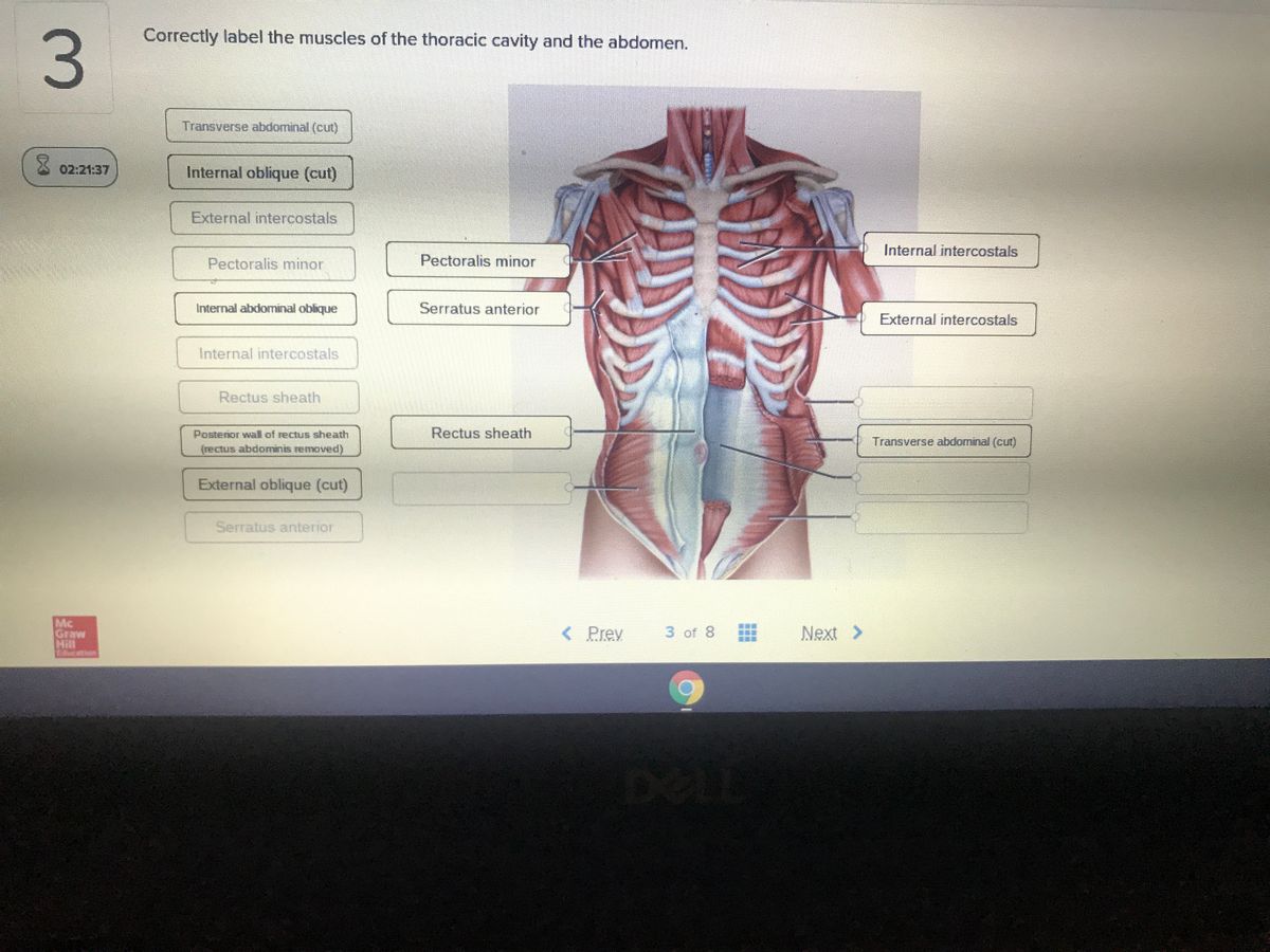
Answered: Correctly label the muscles of the… | bartleby
Thorax: Anatomy, wall, cavity, organs & neurovasculature | Kenhub
The Pleurae - Visceral - Parietal - TeachMeAnatomy
3: The Thorax | Pocket Dentistry

Dorsal and Ventral Body Cavities

Chest Cavity

19.5 Heart Position Within the Thoracic Cavity. (a) The heart ...

Body Cavities - Body Cavities-Internal Chambers Body cavities ...
Thorax: Anatomy, wall, cavity, organs & neurovasculature | Kenhub
Solved Award: 0.76 points Label the structures of the | Chegg.com
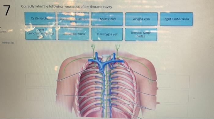
Solved Correctly label the following lymphatics of the ...
Post a Comment for "43 label thoracic cavity"