43 label the internal anatomy of the heart
mammalian heart anatomy worksheet internal Heart labeling (internal). Anatomy heart blank diagram human label unlabeled science diagrams. Free unlabelled diagram of the heart, download free unlabelled diagram ... human labeled draw well sectional sketch structure easy section ncert biology flow class cross blood labelled drawing label. Heart anatomy worksheet diagram bio system ... Anatomy of the Human Heart the outer parietal pericardium. The internal anatomy of the heart reveals four chambers composed of cardiac muscle or myocardium. The two upper.
Color Diagrams of Insect Organs and Internal Structures 17/01/2019 · Perforations in the heart wall, called ostia, allow hemolymph to enter the chambers from the body cavity. Muscle contractions push the hemolymph from one chamber to the next, moving it forward toward the thorax and head. In the thorax, the blood vessel is not chambered. Like an aorta, the vessel simply directs the flow of hemolymph to the head. Insect blood is only …

Label the internal anatomy of the heart
A&P - Anatomy & Physiology: The Unity of Form and Function ... Correctly label the following gross anatomy of the hypothalamus and pituitary glands. Identify the hormone abbreviations and classify them by their main target organs. Correctly label the following structures related to the parathyroid gland. Anatomy and Physiology 2e - 2e - Open Textbook Library Anatomy and Physiology 2e is developed to meet the scope and sequence for a two-semester human anatomy and physiology course for life science and allied health majors. The book is organized by body systems. The revision focuses on inclusive and equitable instruction and includes new student support. Illustrations have been extensively revised to be clearer and … How to Draw the Internal Structure of the Heart (with Pictures) - wikiHow 1. To find a good diagram, go to Google Images, and type in "The Internal Structure of the Human Heart". Find an image that displays the entire heart, and click on it to enlarge it. 2. Find a piece of paper and something to draw with. Start with the pulmonary veins.
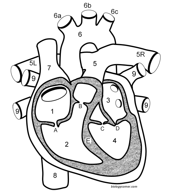
Label the internal anatomy of the heart. Learn the Anatomy of the Heart - The Biology Corner The heart has four chambers, and most diagrams will show the heart as it is viewed from the ventral side. This means that as you look at the heart, the left side refers to the "patient's" left side and not your left side. **For each of the numbers described below, LABEL on the heart diagram.**. Blood that has traveled through the body supplying ... Ch. 19 Circulatory System- heart Flashcards | Quizlet Place the labels in order denoting the flow of blood through the pulmonary circuit beginning with the right atrium and ending in the left atrioventricular valve. The first and last structures are given. Right atrium 1. tricuspid valve 2. right ventricle 3. pulmonary valve 4. pulmonary trunk 5. pulmonary artery 6. lungs 7. pulmonary vein Organ Systems of the Human Body Worksheets Get the basics of human anatomy right with this chart. Label the Organs in the Human Body Once armed with the knowledge of the vital organs and their position in the human body, check if 3rd grade and 4th grade kids can identify and label the internal human organs like the lungs, liver, and more. blank heart diagram worksheet This Is Awesome! Human Anatomy Labeling Worksheets Human Body . worksheets human body anatomy labeling heart. Free Unlabelled Diagram Of The Heart, Download Free Unlabelled Diagram clipart-library.com. heart diagram human parts anatomy unlabeled labeled unlabelled clipart cliparts grade nahar nefarious library anatomical bio
Label the Heart - The Biology Corner Shows a picture of a heart with letters and blanks for practice with labeling the parts of the heart and tracing the flow of blood within the heart. Human Heart (Anatomy): Diagram, Function, Chambers, Location in Body Chambers of the Heart The heart is a muscular organ about the size of a fist, located just behind and slightly left of the breastbone. The heart pumps blood through the network of arteries and... Heart Anatomy: Labeled Diagram, Structures, Blood Flow ... - EZmed Feb 24, 2022 ... Function and anatomy of the heart made easy using labeled diagrams of cardiac structures and blood flow through the atria, ventricles, ... correctly label the following internal anatomy of the heart ... Epicardium is the part of the heart that connects the myocardium and the endocardium. the epicardial wall of the left side of the heart is made of endocardium. The Epicardium is the internal structure of the heart. It is the first layer of the heart wall and is comprised of the endocardium. It is made of endocardium.
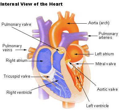
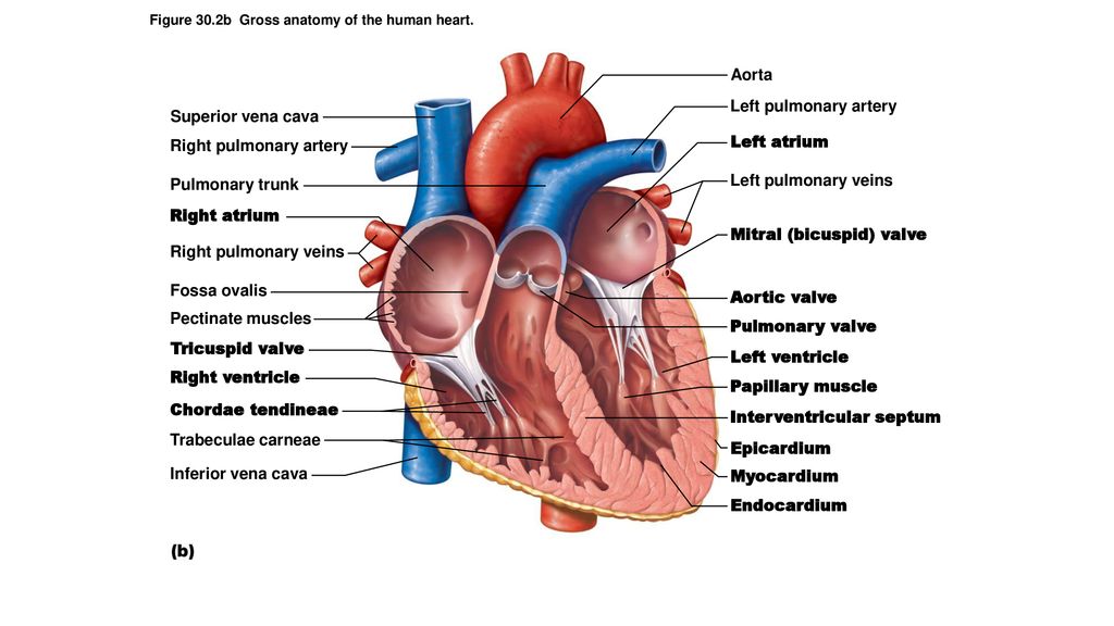
OpenStax AnatPhys fig.19.9 - Internal Anatomy of the Heart Internal Structures of the Heart. This anterior view of the heart shows the four chambers, the major vessels and their early branches, as well as the valves ... Anatomy and Physiology 2e - 2e - Open Textbook Library Anatomy and Physiology 2e is developed to meet the scope and sequence for a two-semester human anatomy and physiology course for life science and allied health majors. The book is organized by body systems. The revision focuses on inclusive and equitable instruction and includes new student support. Illustrations have been extensively revised to be clearer and more inclusive. The web-based ... Correctly Label The Following Internal Anatomy Of The Heart When you study the anatomy of the heart, you will see that it has three main anatomical features. Among them are the aorta, the vena cava, and the pulmonary veins. The heart is made of tissue. It needs nutrients and oxygen. The chambers of the heart are filled with blood. However, the heart does not receive nourishment from the blood. Label the heart — Science Learning Hub Label the heart Interactive Add to collection In this interactive, you can label parts of the human heart. Drag and drop the text labels onto the boxes next to the diagram. Selecting or hovering over a box will highlight each area in the diagram. pulmonary vein semilunar valve right ventricle right atrium vena cava left atrium pulmonary artery
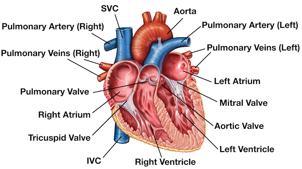
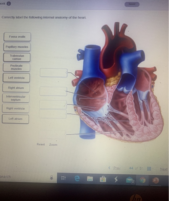
Correctly label the following internal anatomy of the heart. Left pulmonary veins Left ventricle
Layers of the heart: Epicardium, myocardium, endocardium - Kenhub The myocardium is functionally the main constituent of the heart and the thickest layer of all three heart layers. It is a muscle layer that enables heart contractions. Histologically, the myocardium is comprised of cardiomyocytes.Cardiomyocytes have a single nucleus in the center of the cell, which helps to distinguish them from skeletal muscle cells that have multiple nuclei dispersed in the ...
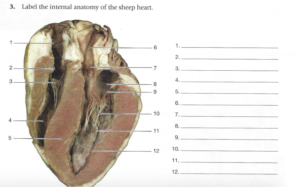
Solved 3. Label the internal anatomy of the sheep heart. 6 ...
Heart Anatomy Labeling Game - PurposeGames.com About this Quiz This is an online quiz called Heart Anatomy Labeling Game There is a printable worksheet available for download here so you can take the quiz with pen and paper. Your Skills & Rank Total Points 0 Get started! Today's Rank -- 0 Today 's Points One of us! Game Points 19 You need to get 100% to score the 19 points available

Atrium (heart) - Wikipedia
internal structure of heart diagram heart structure internal external human circulation science blood. Heart - Anterior Gross Anatomy . anatomy heart anterior gross quiz external purposegames game. Structure Of The Human Heart - YouTube . heart structure human diagram sketch name label parts clipart clip library. Notes: Heart And Circulatory ...

51,449 Human Heart Anatomy Stock Photos and Images - 123RF
Anatomy & Physiology: The Unity of Form and Function - Quizlet Each label contains a hormone and resulting internal body condition. Drag each label to identify whether the condition would be caused by hyposecretion or hypersecretion of the given hormone. HYPERSECRETION • Epinephrine: HYPERSECRETION causes persistently high blood pressure • Estrogen: (in men) HYPERSECRETION causes development of breasts • Follicle stimulating …

The Help of a Well-labeled Diagram Describes the Internal ...
anatomy of the heart valves heart anatomy models chambers biologycorner labeled internal label vessels 3d pulmonary cardiac publisher follow google veins circulatory. Clinical Anatomy Of The Aortic Root | Heart heart.bmj.com. aortic root heart anatomy valve figure bmj coronary pulmonary powerpoint tab open clinical. Anatomy Of The Human Heart - Physiopedia ...

STRUCTURE OF THE INTERNAL HEART. | Biology
Human Anatomy Lab Manual - Open Textbook Library 08/12/2019 · This is a lab manual for a college-level human anatomy course. Mastery of anatomy requires a fair amount of memorization and recall skills. The activities in this manual encourage students to engage with new vocabulary in many ways, including grouping key terms, matching terms to structures, recalling definitions, and written exercises. Most of the activities in this …

Pin on Paramedic Study Guide
Organ Systems of the Human Body Worksheets - Math … This show-and-tell internal organs of the human body printable chart displays the major organs like heart, lungs, liver, etc., and their location in the human body. Get the basics of human anatomy right with this chart. Label the Organs in the Human Body. Once armed with the knowledge of the vital organs and their position in the human body, check if 3rd grade and 4th …

File:Heart diagram-en.svg - Wikimedia Commons
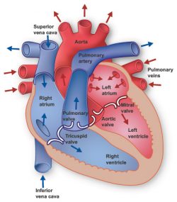
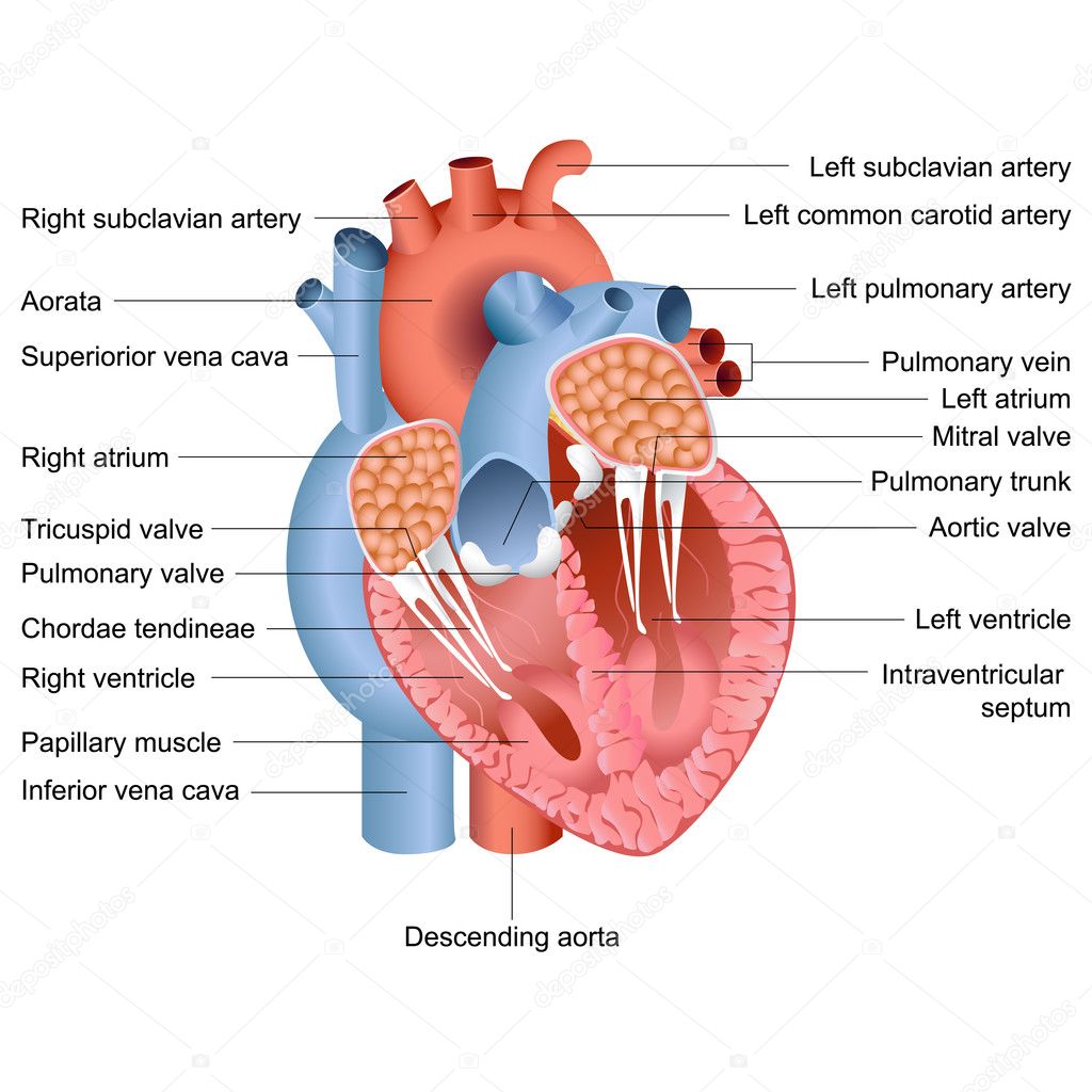
PDF Internal Anatomy of Heart - Mrs. Hille's FunZone THE INTERNAL ANATOMY OF THE HEART Instructions: (1.) Read the statements. (2.) Use the statements to help you to label the diagram and answer the questions. RI Statements 1. Inside the heart are four spaces or chambers. Each chamber in the top half of the heart is called an "atrium"; the plural form is "atria." Arrows D and J point to the atria.

3. internal structure of the heart
Cardiovascular system Diagram - Quizlet Image: correctly label the external anatomy of the anterior heart. Correctly label the following ... Image: Label the internal anatomy of the heart.

4,112 Human Heart Diagram Stock Photos, Pictures & Royalty ...
Basic Anatomy of the Heart - Health Encyclopedia - University of ... The adult human heart is about the size of a fist. The heart beats at an average rate of 80 times a minute. That's about 115,000 times in one day, or about 42 million times in a year. In a 70-year lifetime, an average human heart will beat more than 2.5 billion times. The heart works hard even when you are at rest.
17.5: Internal Structures of the Heart - Biology LibreTexts
Heart anatomy: Structure, valves, coronary vessels | Kenhub Heart anatomy Heart valves Blood flow through the heart Coronary circulation Great vessels of the heart Clinical notes Sources + Show all Heart anatomy The heart has five surfaces: base (posterior), diaphragmatic (inferior), sternocostal (anterior), and left and right pulmonary surfaces.
Heart Anatomy: Heart Dissection
Anatomy quizzes - PurposeGames Label the Heart by LMaggieO 1,487,190 plays 21p Image Quiz. ... Anatomy of the Human Heart - Internal Structures by orkide1 123,967 plays 24p Image Quiz.
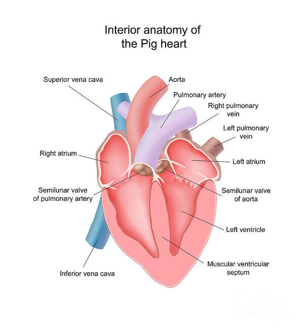
Pig Heart Interior Anatomy Poster by Carlyn Iverson | Fine ...
Label Internal Anatomy of The Heart Diagram | Quizlet Superior vena cava ... Branches of right pulmonary artery ... Aortic semilunar valve ... Right pulmonary veins ... Pulmonary semilunar valve ... Right atrium ... Coronary sinus ... Right atrioventricular canal ... Tricuspid valve ... Papillary muscles ... Right ventricle ... Inferior vena cava ... Aortic arch ... Left pulmonary artery ...

AHCDW15Notes14.pdf - 14. Award: 1.00 point Problems? Adjust ...
Chapter 20-Cardiovascular System Flashcards | Quizlet Correctly label the following internal anatomy of the heart. b Place the labels in order denoting the flow of oxygenated blood through the heart beginning with the vessels that bring blood back to the heart from the lungs. Correctly label the following coronary blood vessels of the heart.

Label the Heart Quiz
Human Anatomy Lab Manual - Open Textbook Library Dec 08, 2019 · This is a lab manual for a college-level human anatomy course. Mastery of anatomy requires a fair amount of memorization and recall skills. The activities in this manual encourage students to engage with new vocabulary in many ways, including grouping key terms, matching terms to structures, recalling definitions, and written exercises. Most of the activities in this manual utilize anatomical ...
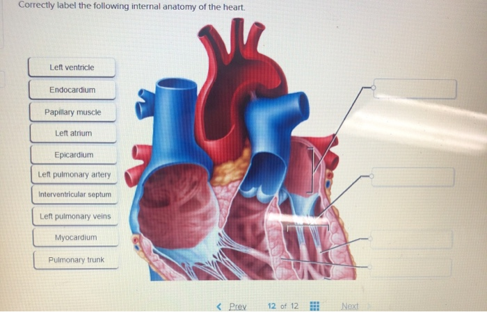
Solved Correctly label the following internal anatomy of the ...
Internal Anatomy of the Heart Flashcards | Quizlet anatomy and physiology Following an injury to a nerve, the muscles it supplies with motor nerve fibers may become paralyzed. How would you explain to a patient the importance of moving the disabled muscles passively or contracting them with electrical stimulation?
Heart Information Center: Heart Anatomy | Texas Heart Institute
Anatomy and Physiology - Wiki - Scioly.org 06/09/2022 · Anatomy and Physiology (previously known as A is for Anatomy (1988-1993) and Anatomy (2007-2015) in Division B) is an event which tests students' knowledge about the anatomy and physiology of various systems in the human body.Division B and Division C will both typically concentrate on three systems, which change each year. Specific topics about …

Pin on Photography
Solved 3. Label the internal anatomy of the sheep heart. 6 2 - Chegg Expert Answer 100% (10 ratings) 1. Right atrium. 2. Pectinate muscle 3. Cusp of tricus … View the full answer Transcribed image text: 3. Label the internal anatomy of the sheep heart. 6 2 7 3. 3 10 7. 8. 9. 10. 5 12 12. Previous question Next question
Heart: Anatomy and Function
Heart: Anatomy and Function - Cleveland Clinic Heart. Your heart is the main organ of your cardiovascular system, a network of blood vessels that pumps blood throughout your body. It also works with other body systems to control your heart rate and blood pressure. Your family history, personal health history and lifestyle all affect how well your heart works. Appointments 800.659.7822.
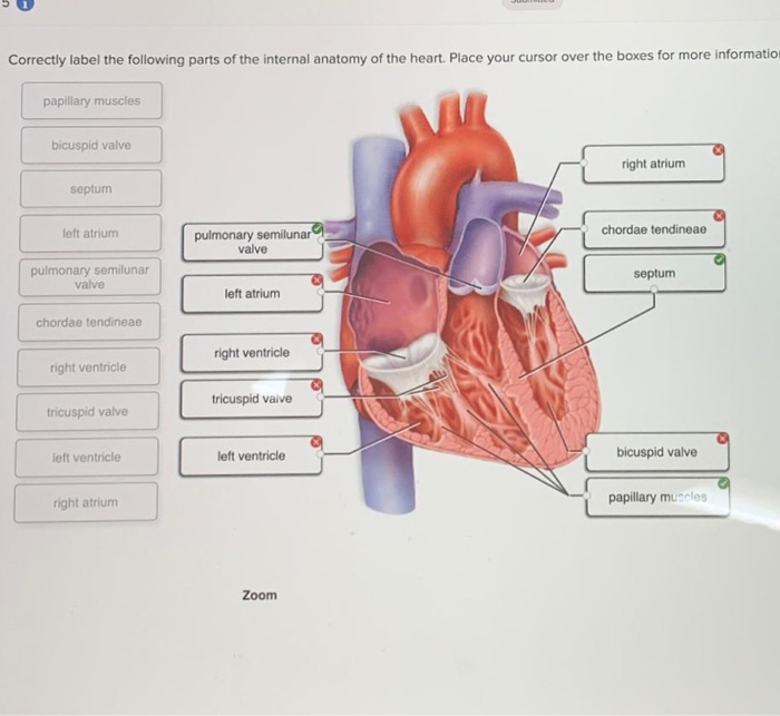
Solved Correctly label the following parts of the internal ...
Internal Structure of the Heart | Contemporary Health Issues In order to develop a more precise understanding of cardiac function, it is first necessary to explore the internal anatomical structures in more detail.
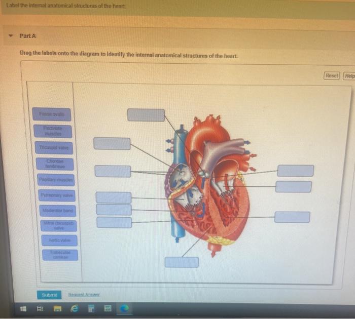
Solved Label the internal anatomical structures of the ...
Heart Information Center: Heart Anatomy | Texas Heart Institute Your heart has 4 chambers. The upper chambers are called the left and right atria, and the lower chambers are called the left and right ventricles. A wall of ...
SEER Training: Structure of the Heart
Chambers of the Heart - Cleveland Clinic Your heart is located under your ribcage just left of your breastbone and between your lungs. The chambers within your heart are arranged in a particular way to allow blood to flow throughout your body. To remember that your atria are the "upper chambers," you can think of them as "above" your ventricles. Both atria and above begin with "a."

Internal Structure of the Heart | Contemporary Health Issues
Anatomy of the Human Heart - Physiopedia Anteriorly: the body of the sternum, and adjoining costal cartilages; left lung, and pleura (apex) · Posteriorly: oesophagus, descending thoracic aorta, azygos, ...
Anatomy of the Human Heart
Anatomy of a referral: Why wait times for specialists are still too ... 30/03/2021 · Anatomy of a referral: Why wait times for specialists are still too long . 61 Comments. Share on: Nancy is “ beyond frustrated” with how long her family has had to wait to see specialists. She has been waiting 16 months to see a rheumatologist, and has been told there’s a two-year wait to a pain management centre to help with her chronic pain, despite the …
Learn the Anatomy of the Heart
Devin Townsend discography - Wikipedia Devin Townsend is a Canadian musician, songwriter, and record producer.. After launching his musical career in 1993 with singing effort on Vai's Sex & Religion and subsequent tour, Townsend released the album Heavy as a Really Heavy Thing under the pseudonym Strapping Young Lad, in 1995. His debut solo release was 1996's Punky Brüster – Cooked on Phonics.

Heart anatomy - Frontal section | Medical anatomy, Heart ...
The Anatomy of the Heart, Its Structures, and Functions - ThoughtCo The heart is the organ that helps supply blood and oxygen to all parts of the body. It is divided by a partition (or septum) into two halves. The halves are, in turn, divided into four chambers. The heart is situated within the chest cavity and surrounded by a fluid-filled sac called the pericardium. This amazing muscle produces electrical ...

CH. 20 Assessment Flashcards | Quizlet
Anatomy and Physiology - Wiki - Scioly.org Sep 06, 2022 · Anatomy and Physiology (previously known as A is for Anatomy (1988-1993) and Anatomy (2007-2015) in Division B) is an event which tests students' knowledge about the anatomy and physiology of various systems in the human body. Division B and Division C will both typically concentrate on three systems, which change each year. Specific topics ...
Heart (right and left atrium): Anatomy and function | Kenhub
Label Heart Interior Anatomy Diagram This cycle is then repeated. Every day, the heart pumps about 2,000 gallons (7,600 liters) of blood, beating about 100,000 times. Label the heart anatomy ...

Internal Anatomy of the Heart Diagram | Quizlet
Chapter 22 Heart Flashcards | Quizlet Label the order that blood flows through in the heart, using the arrows as guides. Label the components of the heart wall. Label the components of the heart as seen from a posterior view. Label the major coronary veins. Label the components of the conduction system. Label the structures of the heart.
Lesson | The Heart - External Structure | Encounter Edu
Anatomy of Insect Organs and Internal Structures Jan 17, 2019 · A single blood vessel runs along the dorsal side of the insect, from the head to the abdomen. In the abdomen, the vessel divides into chambers and functions as the insect heart. Perforations in the heart wall, called ostia, allow hemolymph to enter the chambers from the body cavity.

Heart cross section labeled. Cross section of human heart ...
Home Page: Annals of Vascular Surgery 28/09/2022 · New Journal Launched! Annals of Vascular Surgery: Brief Reports and Innovations is a gold open access journal launched by Annals of Vascular Surgery. The new surgical journal seeks high-quality case reports, small case series, novel techniques, and innovations in all aspects of vascular disease, including arterial and venous pathology, trauma, arteriovenous …
Heart Anatomy: Labeled Diagram, Structures, Blood Flow ...
heart internal anatomy lung labeled respiratory anatomy system larynx labels label lungs diagram labeling using projects cartilage. Label The Heart Quiz . heart human diagram interior game unlabelled label quiz games purposegames statistics. External Gross Anatomy Of The Heart: Anterior View . heart anatomy external anterior ...

3. internal structure of the heart
anatomy of the heart answers heart anatomy internal structures human quiz How Would You Label The Structures (both External And Internal) Of A heart pig anatomy dissection anterior label pigs human dissected sheep labeled lab system external internal circulatory lung structures cow labled Interatrial Septum. Causes, Symptoms, Treatment Interatrial Septum

Heart Anatomy | Anatomy and Physiology II
Anatomy quizzes - PurposeGames Label the Skeleton by Mr. Shumaker 940,546 plays 25p Image Quiz. Anterior Skull Bones by mcscole 602,371 plays 15p Image Quiz. The Plant Cell by PieroTheCooliest 481,423 plays 13p Image Quiz. brain anatomy EC by mkellner24 439,293 plays 10p Image Quiz. Basic Brain Anatomy by chwchang 312,909 plays 14p Image Quiz. CARPAL BONES by zookadoo …
Vector Heart Anatomy Stock Vector Image by ©stockshoppe #10376381
Human Heart - Diagram and Anatomy of the Heart - Innerbody Because the heart points to the left, about 2/3 of the heart's mass is found on the left side of the body and the other 1/3 is on the right. Anatomy of the Heart Pericardium. The heart sits within a fluid-filled cavity called the pericardial cavity. The walls and lining of the pericardial cavity are a special membrane known as the pericardium.
Solved ment Correctly label the following internal anatomy ...
19.1 Heart Anatomy - Anatomy and Physiology 2e | OpenStax Location of the Heart. The human heart is located within the thoracic cavity, medially between the lungs in the space known as the mediastinum. Figure 19.2 shows the position of the heart within the thoracic cavity. Within the mediastinum, the heart is separated from the other mediastinal structures by a tough membrane known as the pericardium, or pericardial sac, and sits in its own space ...
Lab Practical Study Guide - ppt download
How to Draw the Internal Structure of the Heart (with Pictures) - wikiHow 1. To find a good diagram, go to Google Images, and type in "The Internal Structure of the Human Heart". Find an image that displays the entire heart, and click on it to enlarge it. 2. Find a piece of paper and something to draw with. Start with the pulmonary veins.

A&P - Anatomy & Physiology: The Unity of Form and Function ...
Anatomy and Physiology 2e - 2e - Open Textbook Library Anatomy and Physiology 2e is developed to meet the scope and sequence for a two-semester human anatomy and physiology course for life science and allied health majors. The book is organized by body systems. The revision focuses on inclusive and equitable instruction and includes new student support. Illustrations have been extensively revised to be clearer and …
Medical Internal Organs Body Part Nervous System Anatomy ...
A&P - Anatomy & Physiology: The Unity of Form and Function ... Correctly label the following gross anatomy of the hypothalamus and pituitary glands. Identify the hormone abbreviations and classify them by their main target organs. Correctly label the following structures related to the parathyroid gland.

Chapter 20-Cardiovascular System Flashcards | Quizlet
Solved] Label the structures indicated on this anterior view ...
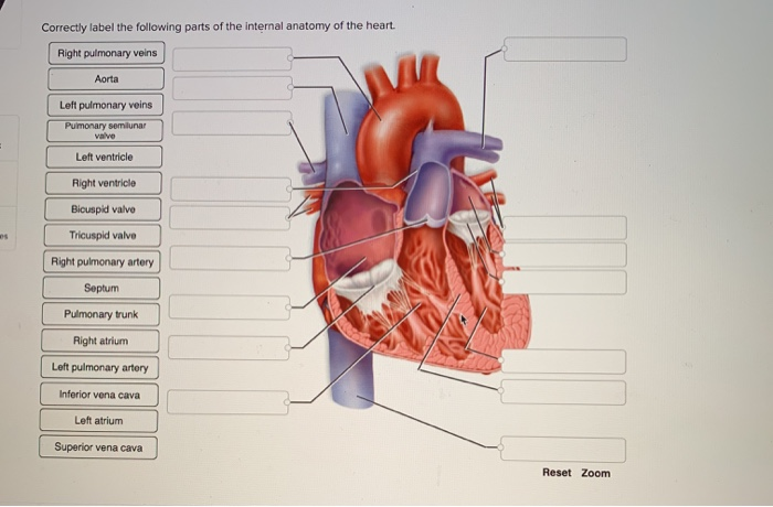
Solved Correctly label the following parts of the internal ...
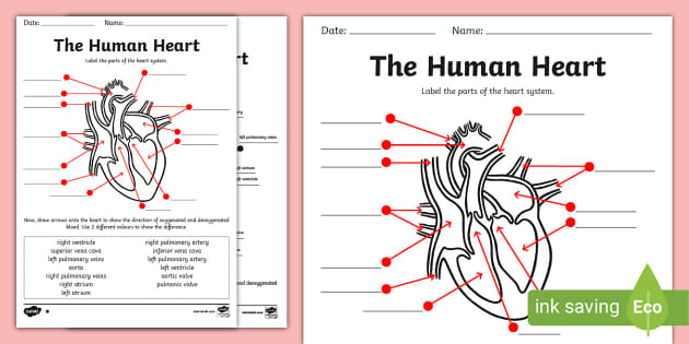
Simple Heart Diagram with Labels Activity - Human Biology
Post a Comment for "43 label the internal anatomy of the heart"